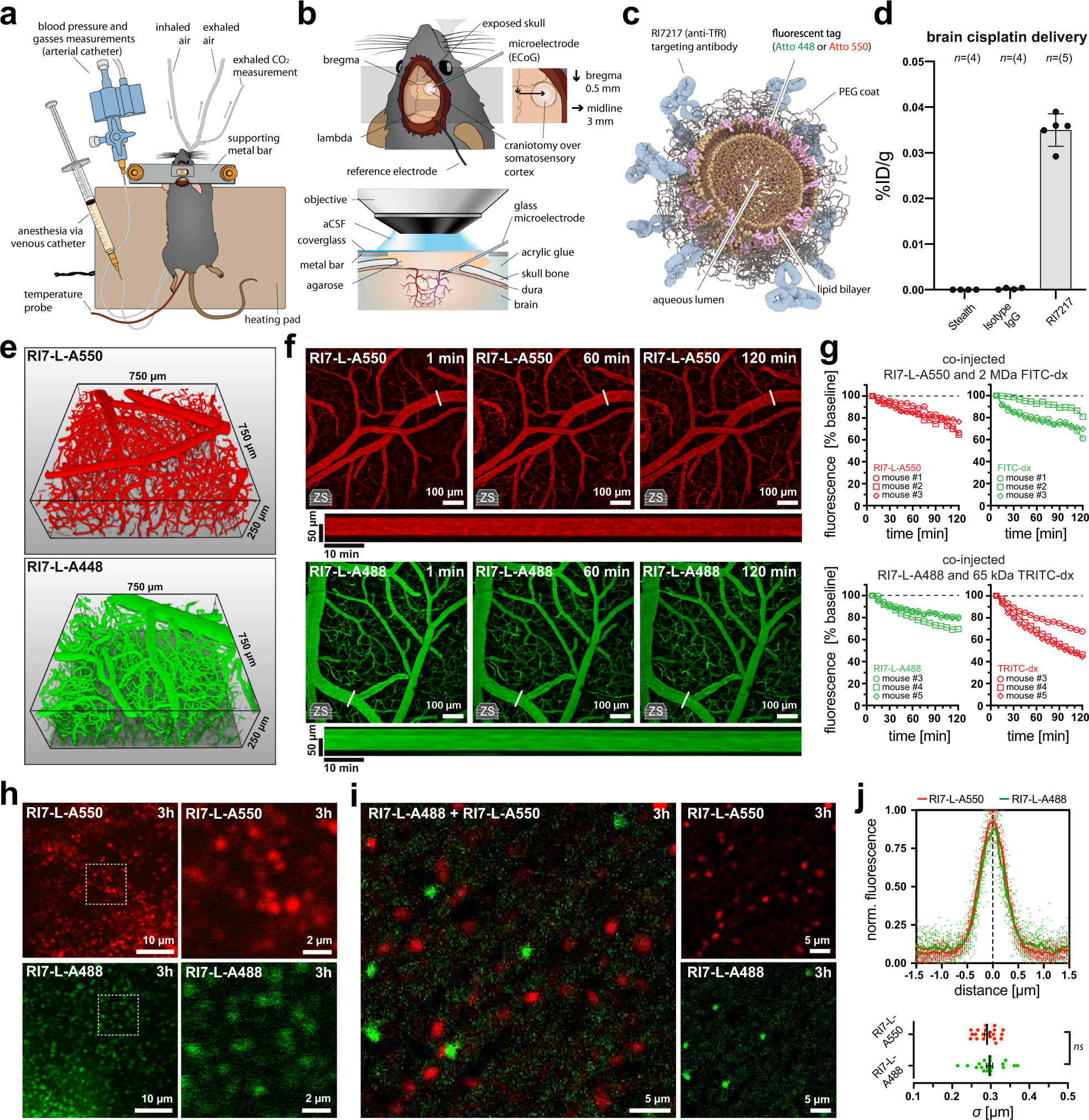
Figure 1 from Brain surface temperature under a craniotomy.
Por um escritor misterioso
Last updated 29 julho 2024

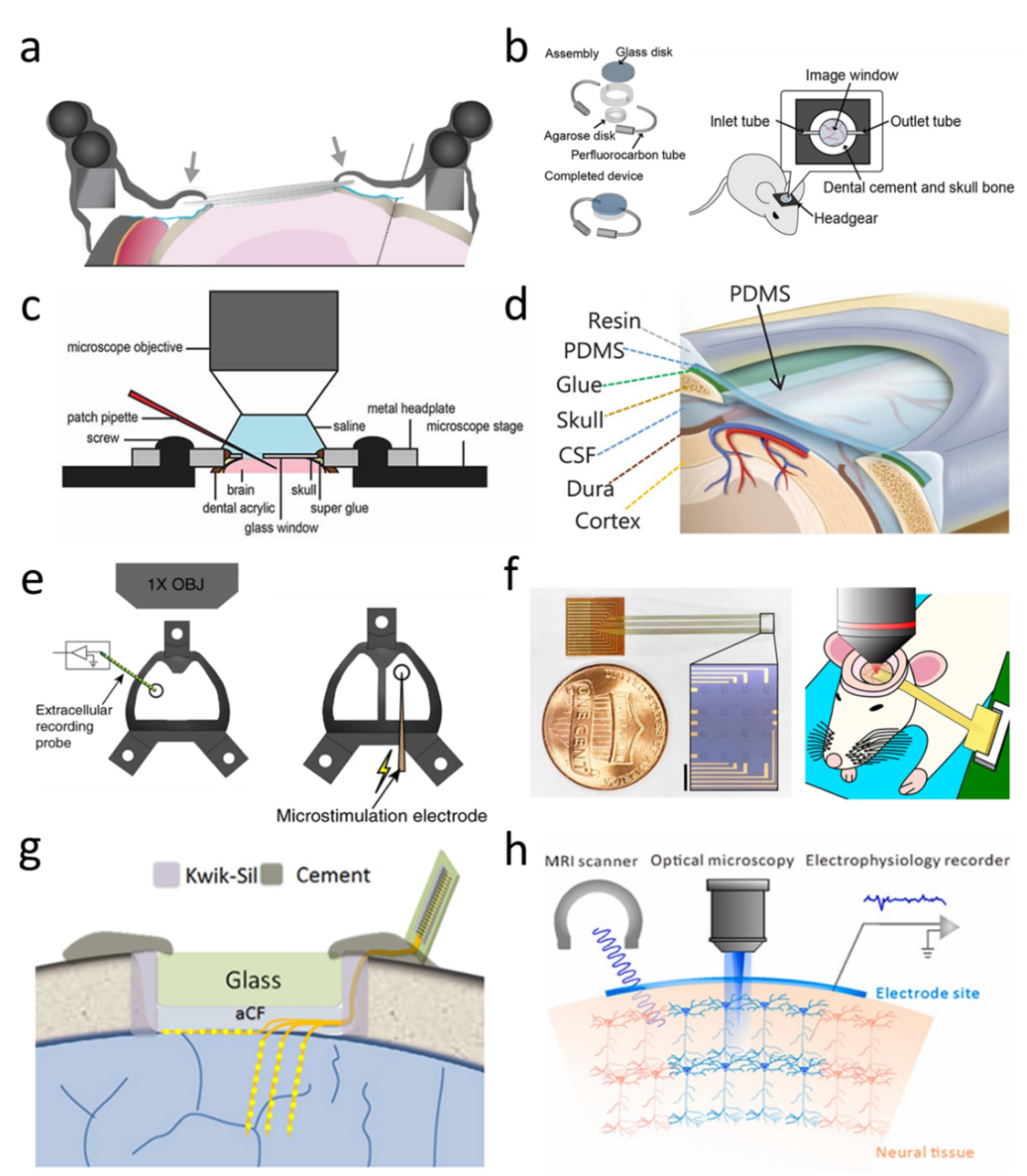
Fig. 1. Rapid cooling of the brain surface in an in vivo mouse preparation. A: schematic representation of a cranial window during recording of temperature and single-cell activity in the anesthetized mouse. The main potential routes of heat transfer are indicated. B: brain surface temperature measured with the thermocouple during replacement of the artificial cerebrospinal fluid (ACSF) with fresh ACSF warmed to 38°C. ACSF was replaced twice, indicated by the arrowheads. - "Brain surface temperature under a craniotomy."
Brain Sciences, Free Full-Text

Applications of flexible electronics related to cardiocerebral vascular system - ScienceDirect

Differential Recovery of Submodality Touch Neurons and Interareal Communication in Sensory Input-Deprived Area 3b and S2 Cortices
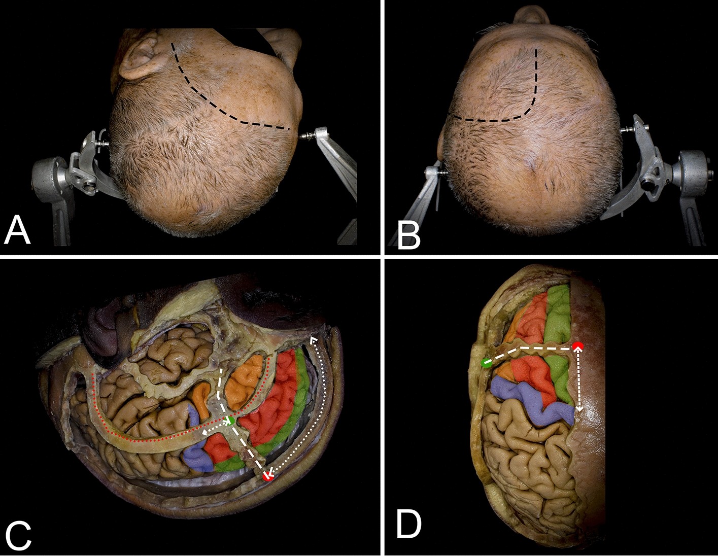
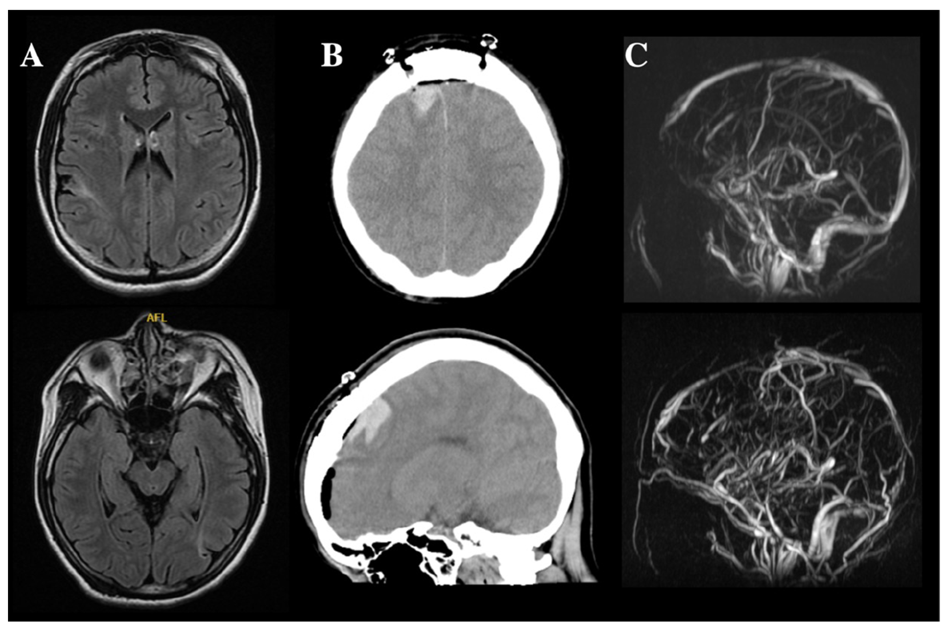
Cortical and white matter anatomy relevant for the lateral and superior approaches to resect intraaxial lesions within the frontal lobe
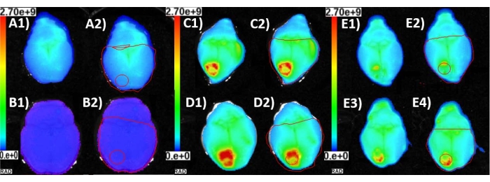
Assessment of Thermal Damage from Robot-Drilled Craniotomy for Cranial Window Surgery in Mice

Data collection and craniotomy. Left: The infrared camera setup is

Temporal/Subtemporal Craniotomy
Electronics, Free Full-Text
Quantitative third-harmonic generation imaging of mouse visual cortex areas reveals correlations between functional maps and structural substrates
Post-capillary venules are the key locus for transcytosis-mediated brain delivery of therapeutic nanoparticles

Transection of the Superior Sagittal Sinus Enables Bilateral Access to the Rodent Midline Brain Structures
Recomendado para você
-
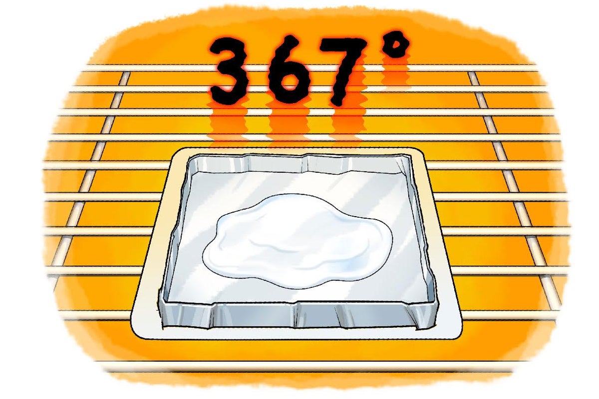
 BRAIN TEST NÍVEL 367 EM PORTUGUÊS29 julho 2024
 -
 Brain Test Level 367 It's cold the fireplace needs more fire in 202329 julho 2024
 -
 Brain Test Level 367 answer/solution. #shorts #braintest29 julho 2024
 -
 Brain Test Nivel 367 - Quiere tener grandes músculos29 julho 2024
 -
 Easy Game Level 367 Find a drag store Answer - Daze Puzzle29 julho 2024
 -
 SumDivide-best FREE block blasting puzzle game to test your brain::Appstore for Android29 julho 2024
 -
 Test your brain Cold Spring Harbor Laboratory29 julho 2024
 -
 Making a Sugar Thermometer29 julho 2024
 -
UTRGV Office For Sustainability - The Rio Grande Valley - Society For Neuroscience- Chapter (RGV-SFN-C) is organizing several events in its mission of promoting: Outreach, Education, Research in the Neuroscience29 julho 2024
-
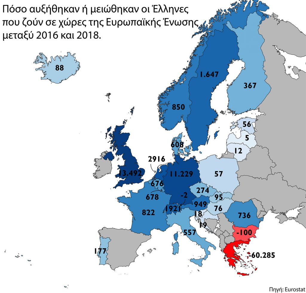 Από το Brain Drain στο Brain Gain: Έτσι μπορεί να αναστραφεί το φαινόμενο (fortunegreece.gr)29 julho 2024

você pode gostar
-
 Gacha life Esboços bonitos, Desenhando roupas de anime, Desenhos kawaii29 julho 2024
 -
 Novak Djokovic Beats Andy Murray to Claim Elusive French Open Title - The New York Times29 julho 2024
 -
 GX Studio on X: ⭐ Yasuhisa Kakuja Full Animation ⭐ Here you go29 julho 2024
 -
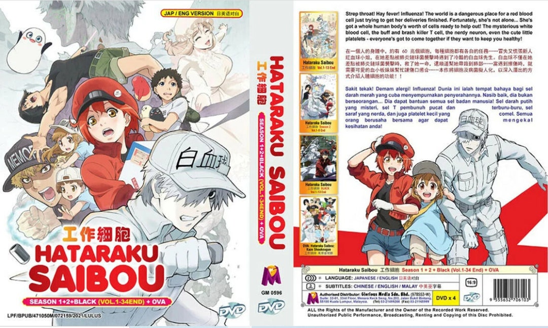 New Set Dvd Anime Hataraku Saibou Season 1 2 Black29 julho 2024
 -
 Topo de bolo Motocross Mulher29 julho 2024
 -
 Free Discord Pfp Maker29 julho 2024
 -
 i.ytimg.com/vi/j2prKN0fmU8/maxresdefault.jpg29 julho 2024
 -
 Inter 2 x 0 Bolívar: gols e melhores lances do jogo pela Libertadores29 julho 2024
 -
 Rblxwild Promo Codes (November 2023)29 julho 2024
 -
 Stream dasdasd music Listen to songs, albums, playlists for free29 julho 2024