Radiological identification and analysis of soft tissue musculoskeletal calcifications, Insights into Imaging
Por um escritor misterioso
Last updated 11 julho 2024
Abstract Musculoskeletal calcifications are frequent on radiographs and sometimes problematic. The goal of this article is to help radiologists to make the correct diagnosis when faced with an extraosseous musculoskeletal calcification. One should first differentiate a calcification from an ossification or a foreign body and then locate the calcification correctly. Each location has a specific short differential diagnosis, with minimal further investigation necessary. Intra-tendon calcifications are most frequently associated with hydroxyapatite deposition disease (HADD). In most cases, intra-articular calcifications are caused by calcium pyrophosphate dihydrate (CPPD) crystal deposition disease. Soft tissue calcification can be caused by secondary tumoural calcinosis from renal insufficiency, or collagen vascular diseases and by vascular calcifications, either arterial or venous (phlebolith). Teaching Points • Calcifications have to be differentiated form ossification and foreign body. • A musculoskeletal MRI study must always be correlated with a radiograph. • The clinical manifestations of calcifications may sometimes mimic septic arthritis or sarcoma. • HADD and CPPD crystal deposition have a distinct appearance on radiograph. • Calcinosis is more frequently caused by chronic renal failure and scleroderma.

Radiological identification and analysis of soft tissue musculoskeletal calcifications. - Abstract - Europe PMC
Calcified or ossified benign soft tissue lesions that may simulate malignancy
Radiological identification and analysis of soft tissue musculoskeletal calcifications, Insights into Imaging

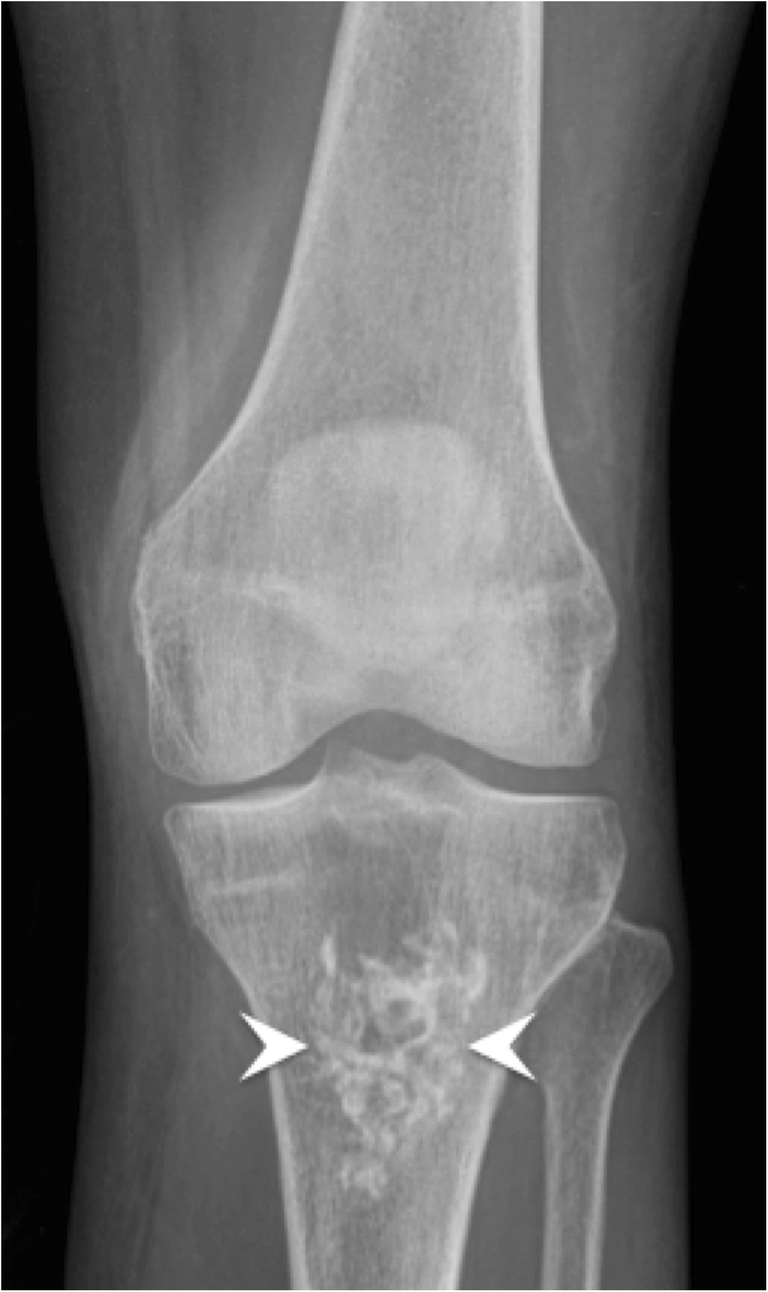
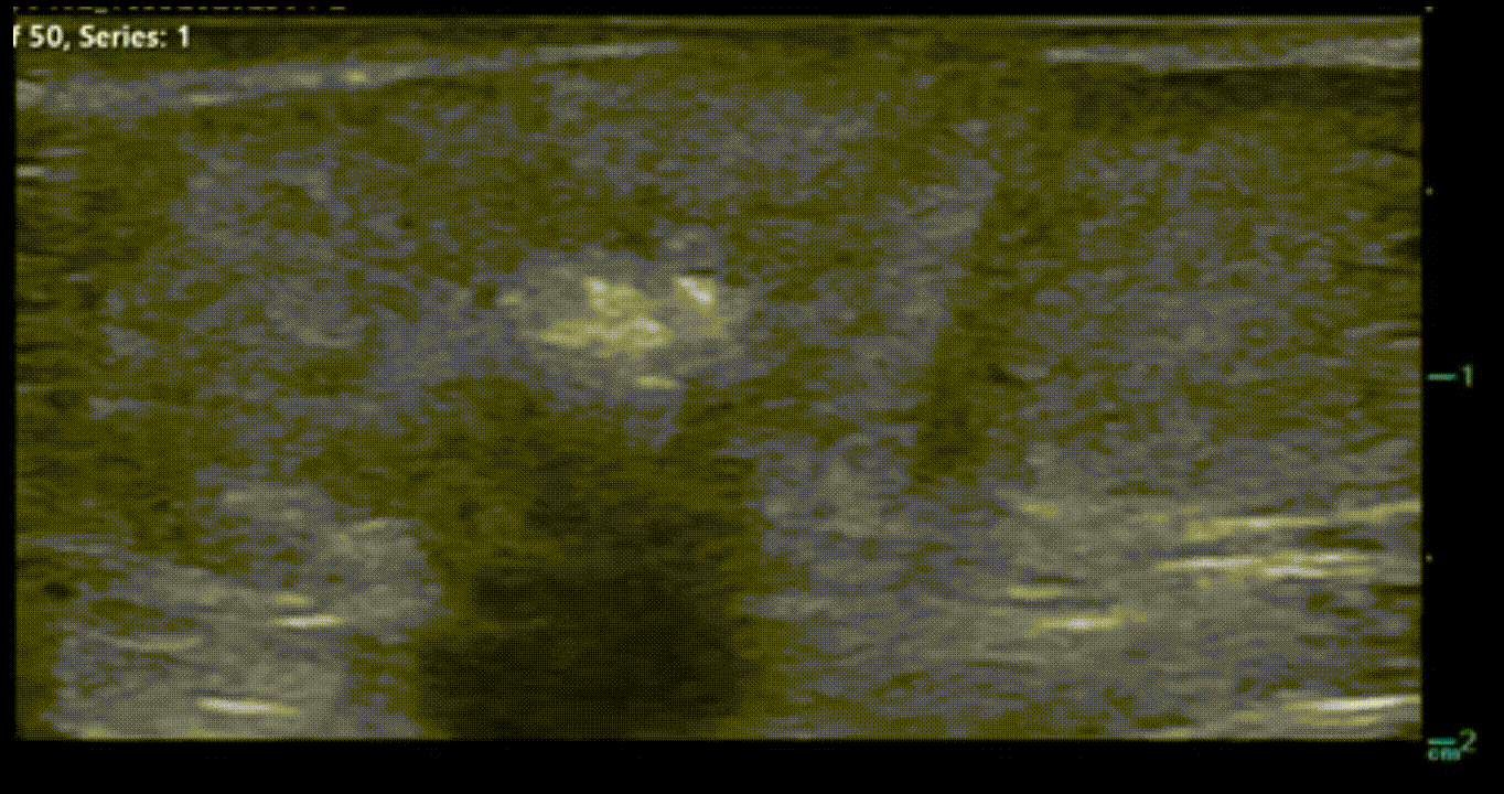
Radiograph of hyaline and fibrocartilage calcification in the hip. The

Soft-Tissue Masses and Masslike Conditions: What Does CT Add to Diagnosis and Management?

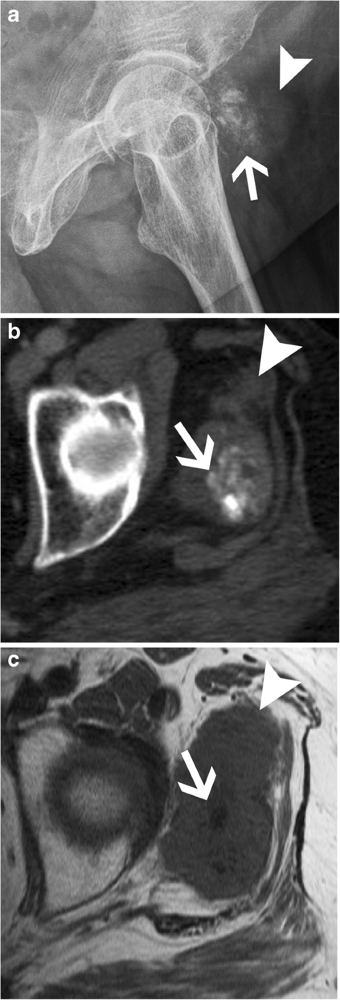
Hydroxyapatite Deposition Disease (HADD) of the Greater Trochanter

Radiological identification and analysis of soft tissue musculoskeletal calcifications. - Abstract - Europe PMC

Ultrasonography of Superficial Soft-Tissue Masses: Society of Radiologists in Ultrasound Consensus Conference Statement
Radiological anatomy: X-ray, CT, MRI
Musculoskeletal Archives - UCSD Ultrasound
Recomendado para você
-
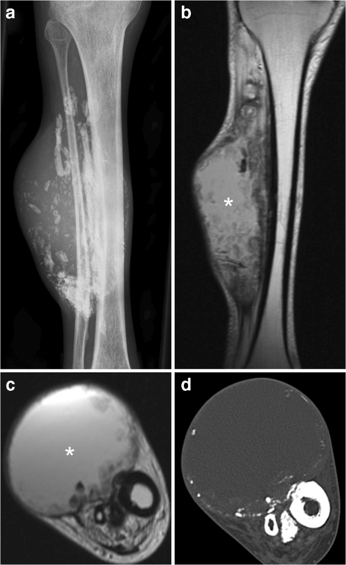
 Lateral radiograph of the left tibia demonstrating the characteristic11 julho 2024
 -
 In the tibia hi-res stock photography and images - Alamy11 julho 2024
 -
 Is there some trick to managing plasma rings? : r/TibiaMMO11 julho 2024
 -
 MMORPG: Rising Force Online, RuneScape, Final Fantasy XI, Tibia, Hero Online, Silkroad Online, Ragnarök Online, The Lord of the Rings Online11 julho 2024
 -
 Tibia Lengthening and Deformity Correction With a Multiplanar External Fixator11 julho 2024
 -
 Loot de 100 Life Rings en Orc Fortress11 julho 2024
 -
 24 inches Real Giraffe Tibia Leg Bone for $99.9911 julho 2024
 -
EPOS™11 julho 2024
-
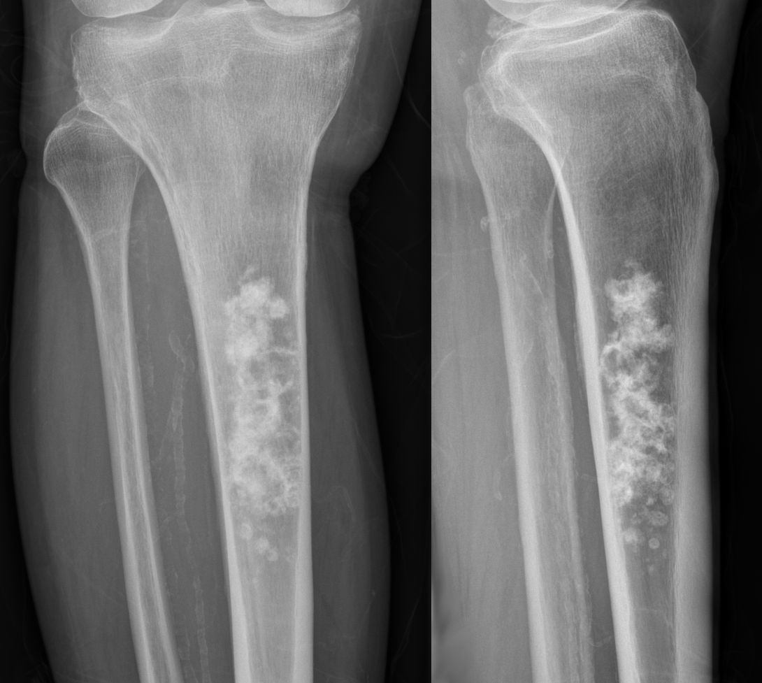 File:Enchondrom Tibia 83jm - Roe ap und seitlich - 001.jpg - Wikimedia Commons11 julho 2024
 -
 Alicorn Ring, TibiaWiki11 julho 2024
você pode gostar
-
 MP investiga jogo em que Bolsonaro mata gays, negros e feministas – Tecnoblog11 julho 2024
 -
 No Friends? Play Solo Chess! SnatchPato on Chess.com11 julho 2024
 -
 bRO changelog May 30, 2023 - Changelog - Divine Pride11 julho 2024
 -
 Tenjho Tenge: Season 1 (2004) — The Movie Database (TMDB)11 julho 2024
 -
 Quebra-Cabeça Grandão - Princesa Disney - 48 Peças - Jak - Ri Happy11 julho 2024
 -
 Uncharted 3: Drake's Deception (PS3)11 julho 2024
 -
Steam Workshop::John Doe Game11 julho 2024
-
 A Slice of Scranton: “The Office” exhibit delivers Dunder Mifflin11 julho 2024
 -
 Making Nezuko in Gacha Club11 julho 2024
 -
 Hear Linkin Park's uncovered Meteora track “Fighting Myself”11 julho 2024